NMMU’s one stop shop for electron microscopy in the Southern Hemisphere
News category: Nanotech
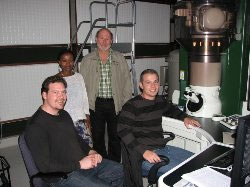
 Professor Jan Neethling (back right) and Dr Jaco Olivier (front right) of NMMU collaborated with Dr Jamie Warner (front left) and Frewyeni Kidane (back left) from Oxford University in a ground breaking study imaging graphene using the HRTEM. Image courtesy of Prof J Neethling
Just two years ago Nelson Mandela Metropolitan University (NMMU) became the first institution in South Africa and Africa to acquire an Ultra-high Resolution Transmission Electron Microscope (U-HRTEM). The U- HRTEM pushes the boundaries of cutting-edge research and training of postgraduate students in advanced electron microscopy and nanoscience.
The JEOL ARM 200F double aberration corrected U-HRTEM at NMMU offers a resolution of a phenomenal 0.08nnm at 200kV. And that level of resolution opens up a whole new world to scientists. The facility, the brain child of NMMU’s Prof Jan Neethling, is literally a game changer in the field of microscopy. Since opening its doors to scientists across SA, the facility has started to tell its own stories, from defects in diamonds, to forensic investigations on the hunt for asbestos, from the mining sector to the study of biological processes, thanks to this new set of eyes.
How does it work? Ultra-high resolution transmission electron microscopy is considered to be an advanced atomic imaging method compared to the traditional TEM available at most universities. These essentially allow for higher resolution imaging of crystallographic structures of samples at a nano and atomic scale. For those in the know, this works by transmitting an electron wave through a sample being studied. This incidental electron wave is scattered at potentials relative to the atoms in the sample, resulting in a change in the phase of the wave. The wave which exits the sample then carries direct, resolved information about the positions of atoms in the sample, the chemical composition of the sample and the nature of the bonds between the atoms in the sample. Following magnification and further phase shifts the final digital image, carrying both a wealth of new information and understanding, is generated.
Over the next few months we will take a look at some of the areas of research where the U-HRTEM is already making substantial impact.
For more information and usage: Prof Jan Neethling: jan.neethling@nmmu.ac.za
Writer: Edith Mshoperi
